I study how bacteria build and rebuild their cell wall, and how the enzymes that reshape peptidoglycan help a cell cope with stress and antibiotics.
About me
I am a bacterial molecular microbiologist. I did my PhD at the CSIR-Centre for Cellular and Molecular Biology (CCMB) in Hyderabad, and I have spent the past three years as a postdoctoral Research Associate at the University of Sheffield, working with collaborators at the Universities of Oxford and Birmingham.
My work is on how bacteria remodel their cell envelope. During my PhD I identified LdtF, the enzyme that releases Braun's lipoprotein from peptidoglycan in E. coli. Afterwards, as a Project Associate at CCMB, I studied how the wall is remodelled during cold shock. In Sheffield I moved to the l,d-transpeptidases of Rhizobium, where we found l-amino acids built into peptidoglycan and an enzyme whose reaction changes with pH.
I combine bacterial genetics, recombinant protein biochemistry, enzyme assays and LC–MS/MS-based peptidoglycomics to study enzyme function, cell-wall architecture and how bacteria adapt, with relevance to antibiotic susceptibility.
Enzymes I have worked on
LdtF
Escherichia coli
A nonconformist enzyme in the LDT family: it cuts Braun's lipoprotein from peptidoglycan.
LdtD
Escherichia coli
Shows how E. coli changes its own jacket during cold shock.
The Rj LDTs are multitasking enzymes that carry out several peptidoglycan-remodelling reactions, breaking the stereotype of one enzyme, one reaction.
LdtRj8 can change the stereochemistry of peptidoglycan.
LdtCd3 supports our finding that l-amino acid incorporation at the C-terminus of the stem peptide is a broader phenomenon.
Research vision
The bacterial cell wall is usually drawn as a fixed scaffold, but its chemistry changes all the time. Enzymes cut, add and exchange residues, and this editing is how a cell keeps its envelope intact in the cold, in acid, under antibiotics and inside a host. I want to know what role cell-wall enzymes play under stress conditions, well enough to predict the wall a cell will build or modify. This matters to me for two reasons. One is to work towards combination therapy against antimicrobial resistance (AMR). The other is that these enzymes are what lets a symbiont remodel its envelope as it settles into its host.
Research domains
My work has two strands: the enzymes that remodel the wall, and the chemistry of the wall itself.
Domain 1, PhD
Enzymes that remodel the cell wall and hold the envelope together
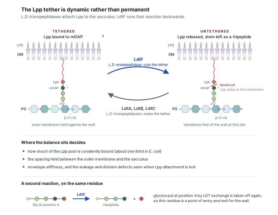
Working model: the tether is reversible. LdtA, LdtB and LdtC attach Lpp to mDAP3; LdtF cuts the same bond. The balance between the two reactions sets how much of the Lpp pool is bound, how far the outer membrane sits from the sacculus, and how well the envelope holds up under stress. The same residue is also where glycine enters the stem and leaves it again. A working model drawn for this page, not a figure from the paper.
Braun's lipoprotein is cut from the cell wall by an l,d-transpeptidase paralog
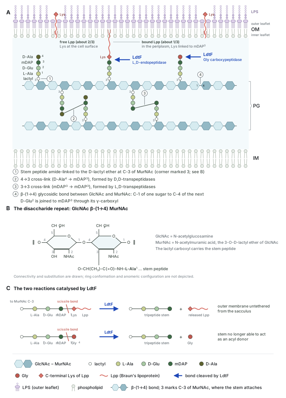
Where LdtF acts in the envelope.(A) Lpp staples the outer membrane to the sacculus: the ε-amino group of its C-terminal lysine is amide-linked to mDAP3 of a stem peptide, while about two-thirds of Lpp stays free with its C-terminus exposed at the surface. Blue arrows mark the two activities of LdtF. (B) The disaccharide repeat, GlcNAc β-(1→4) MurNAc, with the stem peptide carried on the d-lactyl ether at C-3 of MurNAc. (C) Both reactions release a group from mDAP3 and return the stem to a tripeptide: the l,d-endopeptidase reaction untethers the outer membrane, and the glycine-specific carboxypeptidase reaction removes a Gly incorporated at position 4. Drawn for this page; the biology follows Bahadur, Chodisetti & Reddy, PNAS 2021.
In E. coli, Braun's lipoprotein (Lpp) is covalently tethered to peptidoglycan and ties the outer membrane to the cell wall. How that link is broken was unknown. I identified LdtF, a paralog of the l,d-transpeptidases, as the enzyme that cleaves the Lpp–peptidoglycan bond, and showed that it also has carboxypeptidase activity on glycine-containing peptidoglycan.
This gave a function to an orphan member of the LDT family. It also gave the field an enzyme that releases outer-membrane protein from the cell wall, the reverse of the anchoring reaction.
LdtD remodels peptidoglycan in the cold
Nallamotu KC, Bahadur R, Kaul M, Reddy M. Journal of Bacteriology, 2023. doi:10.1128/jb.00382-22
Cells grown at low temperature change the chemistry of their cell wall. We showed that this cold-shock remodelling depends on the l,d-transpeptidase LdtD, which builds 3-3 cross-links in place of the usual 4-3 ones.
This links one enzyme to one environmental stress. It also supports the view that the 3-3 cross-link is a conditional, adaptive feature of the wall, not a constant one.
Domain 2, postdoctoral
Stereochemistry and pH as controls on cell-wall architecture and fitness
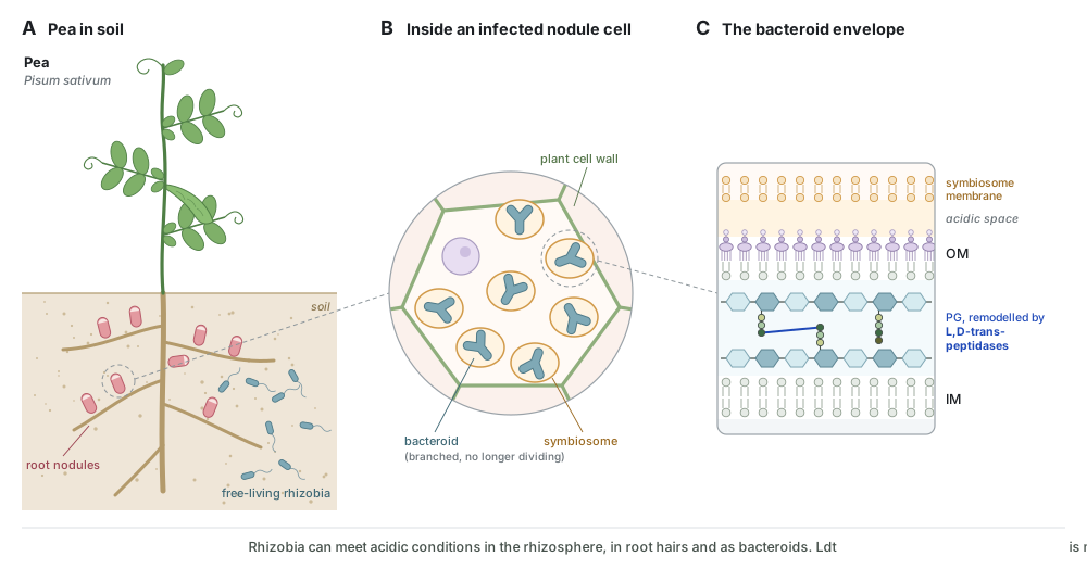
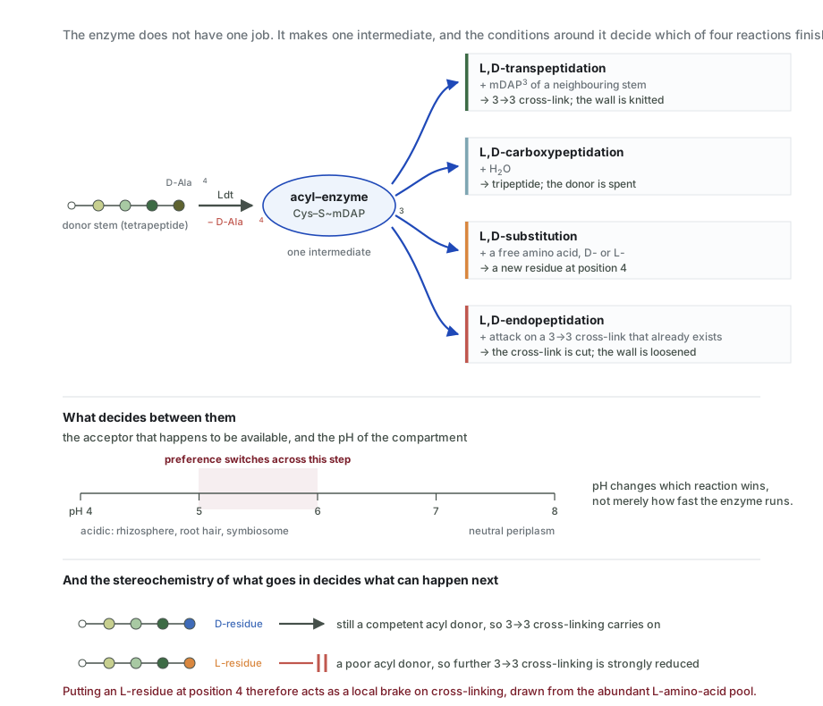
The system these enzymes work in.(A)Rhizobium johnstonii lives free in the soil and forms nodules on the roots of pea (Pisum sativum). (B) Inside an infected nodule cell it becomes a branched bacteroid, each one enclosed in a plant-made symbiosome. (C) The bacteroid envelope, with the peptidoglycan layer that l,d-transpeptidases remodel. These surroundings can be acidic, the range in which LdtRj8 is most active. Whether this remodelling matters in the host is what I want to test. A schematic drawn for this page, not data.Working model: one intermediate, four fates. A single l,d-transpeptidase makes one acyl-enzyme intermediate, and whichever acceptor reaches it first decides the reaction. pH changes which outcome wins, not just how fast the enzyme works. The stereochemistry of the residue installed at position 4 then decides whether the stem can still be used: with an l-residue there, it is a poor donor and further cross-linking slows. A working model drawn for this page, not a figure from the manuscript.
The wall carries both stereochemistries at the end of the stem peptide
Rady BJ*, Bahadur R*, Evans CA, Mesnage S. bioRxiv, 2026. doi:10.64898/2026.06.30.735398. *Equal contribution. Revised manuscript in preparation.
Peptidoglycan is built with d-amino acids at the end of the stem peptide. That stereochemistry is what sets it apart from protein, and what many of its enzymes are assumed to require. Working on Rhizobium johnstonii, we found pairs of muropeptides with the same mass but different retention times. Mass spectrometry could not tell them apart. NMR could: the two differ only in the configuration of the terminal alanine, so a substantial fraction of this wall ends in l-Ala rather than d-Ala.
Screening all 18 l,d-transpeptidases of R. johnstonii in a purpose-built E. coli Lemo21(DE3) Δ6ldt host identified the enzyme responsible. LdtRj8 catalyses every reaction described for the family, and which one it favours shifts with pH. In substitution it accepts all 20 d-amino acids and most l-amino acids, and the same is true of LdtCd3 from Clostridioides difficile, so this is a property of the family, not of a single enzyme.
What it means for fitness
The substitution changes how the wall is built. Replacing the terminal d-Ala with l-Ala cuts subsequent 3-3 cross-linking from 51% to 12% for LdtRj8, and from 33% to 1.2% for LdtCd3. An l-residue at that position is therefore a local brake on cross-linking. It gives the cell a way to control how tightly a patch of wall is cross-linked, using its large pool of l-amino acids instead of scarce d-amino acids. LdtRj8 works best at about pH 5–6. A rhizobium meets that range in the rhizosphere, in root hairs and inside the symbiosome, where it has to remodel its envelope while it colonises the host.
Where I want to take it
I want to take this in two directions. The first is to ask whether l-amino-acid substitution changes fitness in the host, by testing whether a strain that cannot make it is worse at colonisation, at nodulation, or at surviving acid stress. The second is antibacterial. In several pathogens, the 3-3 cross-links made by l,d-transpeptidases are how the cell escapes β-lactams, so an enzyme that can be pushed into substitution instead of cross-linking, either by pH or by the amino acid available to it, is worth testing on that route. The E. coli Δ6ldt strain I built for this work secretes the enzyme into the medium, so it is also a practical way to produce periplasmic proteins that are otherwise hard to express.
Other contributions
Methods development
Developed an ultracentrifuge-free PG isolation workflow in 96-well plates, PG detection by western blotting, in-house mutanolysin production, and paired RP-HPLC and LC–MS methods.
Peptidoglycan hydrolase biology
Characterised the murein endopeptidase MepR, which hydrolyses 3-4 cross-links in anhydromuropeptides (unpublished work).
Bahadur R, Chodisetti PK, Reddy M. Proceedings of the National Academy of Sciences USA, 2021; 118(19): e2101989118. doi:10.1073/pnas.2101989118
Identified LdtF as the enzyme that cuts Braun's lipoprotein from peptidoglycan.
* Co-first authors, contributed equally.
Research experience
2023–2026
Research Associate
University of Sheffield, UK
Investigated PG remodelling and bacterial envelope integrity, with emphasis on the l,d-transpeptidases of Rhizobium johnstonii (formerly R. leguminosarum) 3841.
Characterised 18 putative l,d-transpeptidases and investigated atypical amino-acid incorporation into PG using PG isolation, RP-HPLC, LC–MS/MS, PG-Finder and molecular and biochemical approaches.
Worked on joint projects with groups at the University of Oxford and the University of Birmingham.
2022–2023
Project Associate
CSIR-Centre for Cellular and Molecular Biology (CCMB), India
Investigated LdtD-mediated PG remodelling during cold shock in E. coli.
2016–2022
PhD Researcher
CSIR-Centre for Cellular and Molecular Biology (CCMB), India
Studied PG hydrolases and cell-envelope remodelling in E. coli.
Identified and characterised LdtF and its role in PG–Lpp cross-link cleavage.
2014–2015
Scientific Officer
Bhabha Atomic Research Centre (BARC), Mumbai, India
Education
2022
PhD, Molecular Microbiology
Jawaharlal Nehru University, New Delhi (CSIR-Centre for Cellular and Molecular Biology), India
2012
MSc, Biotechnology
Chhatrapati Shahu Ji Maharaj University, Kanpur, India
2009
BSc
Chhatrapati Shahu Ji Maharaj University, Kanpur, India
Technical expertise
Bacterial genetics
Molecular biology and recombinant DNA
Protein expression and purification (bacterial and insect cell systems)
Peptidoglycan isolation
Peptidoglycomics
LC–MS/MS
RP-HPLC
PG-Finder and computational analysis
Enzyme kinetics
Microscopy
Antibiotic and stress phenotyping
Experimental design and collaboration
Awards, grants and recognition
2025/2026Recognised as outstanding postdoc
2025General Travel Grant, Biochemical Society
2025External Event Grant, Microbiology Society
2023–2026UK Global Talent Visa, Bioscience (UKRI-endorsed route)
2022Best Poster Award, All India Cell Biology Conference
Conferences
20258th Great Wall Symposium, Catania, Sicily, Italy (poster)
20237th Great Wall Symposium, Vila Galé Sintra, Portugal (poster)
202244th All India Cell Biology Conference, University of Kashmir, India (poster)
2018International Congress of Cell Biology, Hyderabad, India (participant)
Contact
I am glad to hear from possible collaborators, and from anyone working on peptidoglycan.